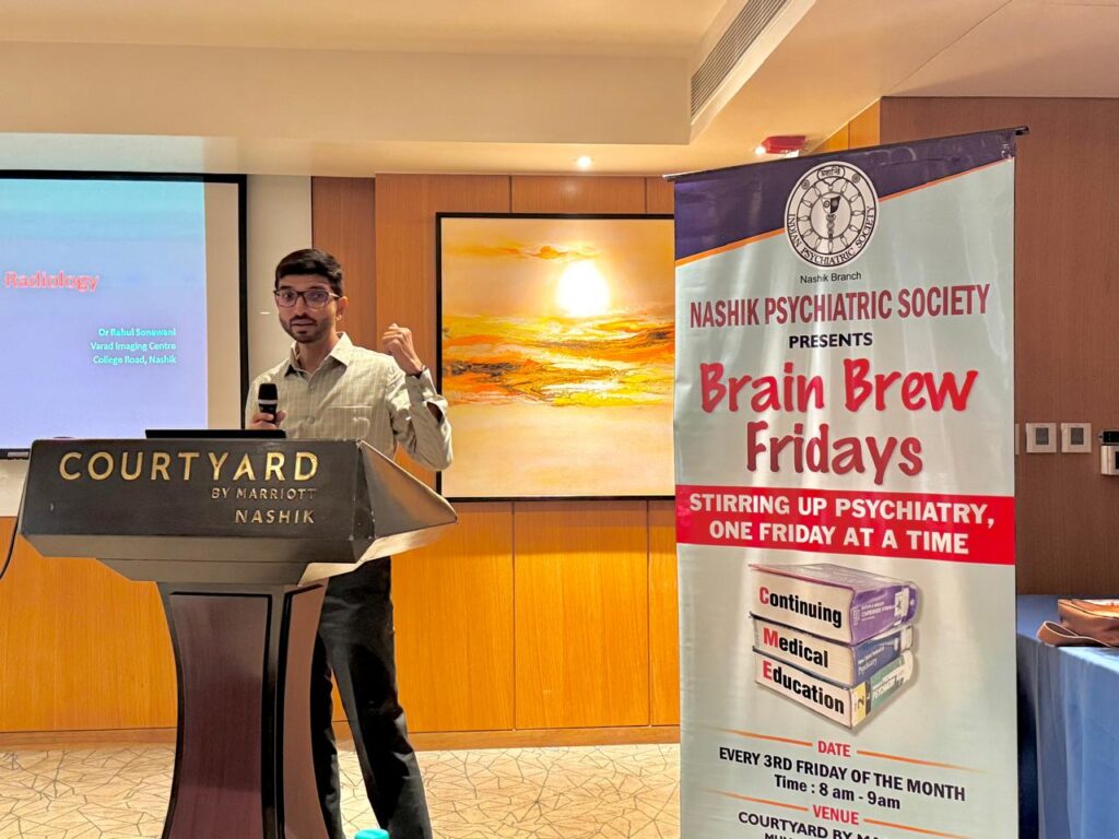
Morning CME 2 - Radiology
Topic: Mind in Radiology
Speaker: Dr. Rahul Soanwani (Consultant Radiologist)
Chairpersons:
1. Dr. BSV Prasad
2. Dr. Devika Patil
Date: 18th July, 2025
Summary:
3 axes
Axial – transverse ,i.e from up to down slices
Sagittal- Right to left(lateral)
Coronal- anterior to posterior
In CT – morphological characteristic seen
In MRI- anatomy seen properly

CT-
- Grey matter seen light coloured
- White matter more grey
- Hyperdense ( solid ) matter appears white like blood, bone ,calcification
- Hypodense appears dark or black like CSF,fluid, abscess,air,fat.
- Fractures seen more clearly in CT
- Also if paralysis or weakness within 15 mins do CT first – helps in diagnosis

MRI-
T1-
- (brother of CT) ,more clarified
- Colour of csf – black
- To identify structure n anatomy
T2-
- better than T1
- CSF- white
- Odema,lesion seen
Flair-
- its T2 weighted image
- More detailed pathology
- Lesions close to csf seen as csf is suppressed
Diffusion weighted –
- To diagnose infarct
Diffusion weighted –
Hypertensive bleed -arterial mainly centrally (thalamus)
Venous bleed - peripheral
1) Epidural bleed – between bone n dura
Not free flowing
Shape concavoconvex ( like lemon)
Cause is fracture
Mostly arterial
2) Subdural -between dura n arachnoid- banana shaped seen
In old age, h/0 fall
3) Subarachnoid – inside brain
4) interventricular bleed

Normal pressure hydrocephalus
- Seen in old age
- Less atrophy with more ventricular dilatation
- Triad of ataxia,cognitive impairmentn urinary incontinence
Epilepsy-
- Temporal region mainly -hippocampus(medial most part)
- Seen in coronal image
- Hippocampus seen lateral to choriod sulcus n below temporal horn
MDD
- Functional MRI, PET more helpful
- MRI- in old age-white dots seen in brain
- In young- subcortical white matter – white dots seen
- Hippocampus atrophy seen
Alzeihmers
- Amyloid in small vessels (surpenginous white dots)
- Atrophy seen
- Slyvian fissure dilated
- Hippocampus atrophy
- Temporal horn dilated

Schizophrenia-
- Thinned cortex
- Enlarged 3rd ,lat ventricle
- Reduced hippocampus
Alcohol abuse
- inappropriate brain atrophy
- more of middle side of thalamus
- 40 years vale ka brain 60 vale jaisa
- widening of cerebellar fossa
- dilatation of sulci

OCD
- Atrophy of caudate nucleus seen


